Yeast
Label-free imaging of unstained yeast — no dyes, no fixation, no fluorescent reporters.
One measurement, four contrast modes
Brightfield for morphology, quantitative phase for dry mass, DIC for fine relief, scattering angle for boundaries and texture — all from one fast, automated measurement, with every cell in the field of view captured at once.
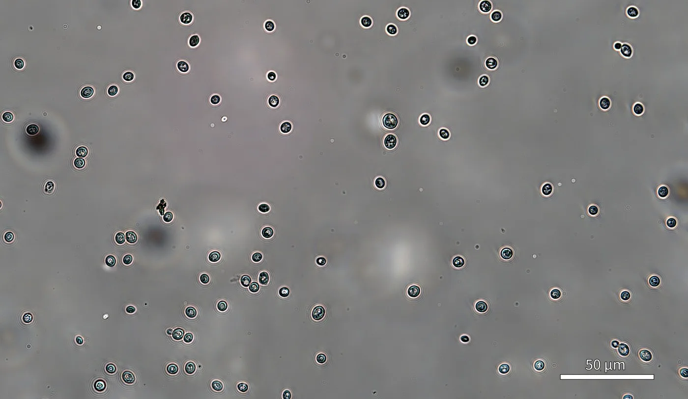
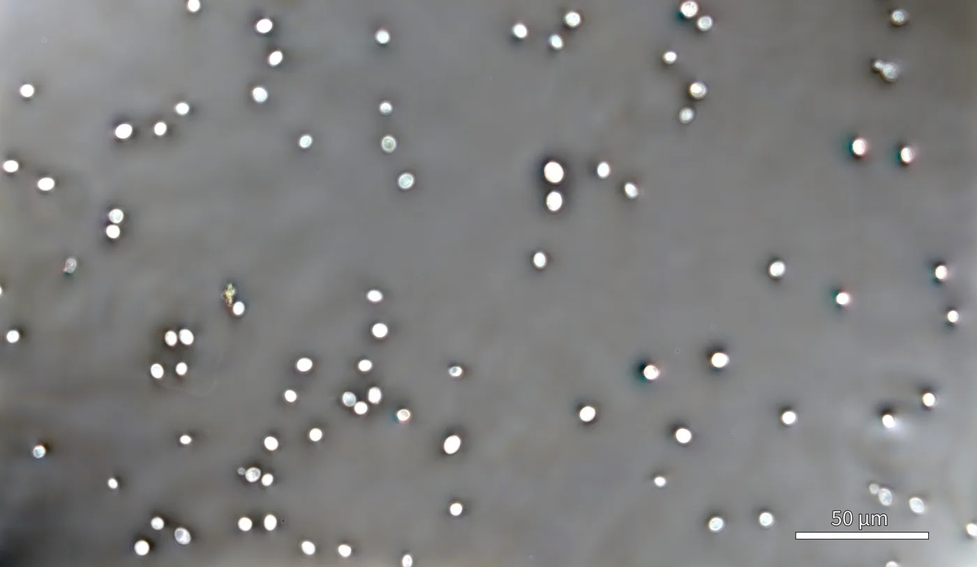
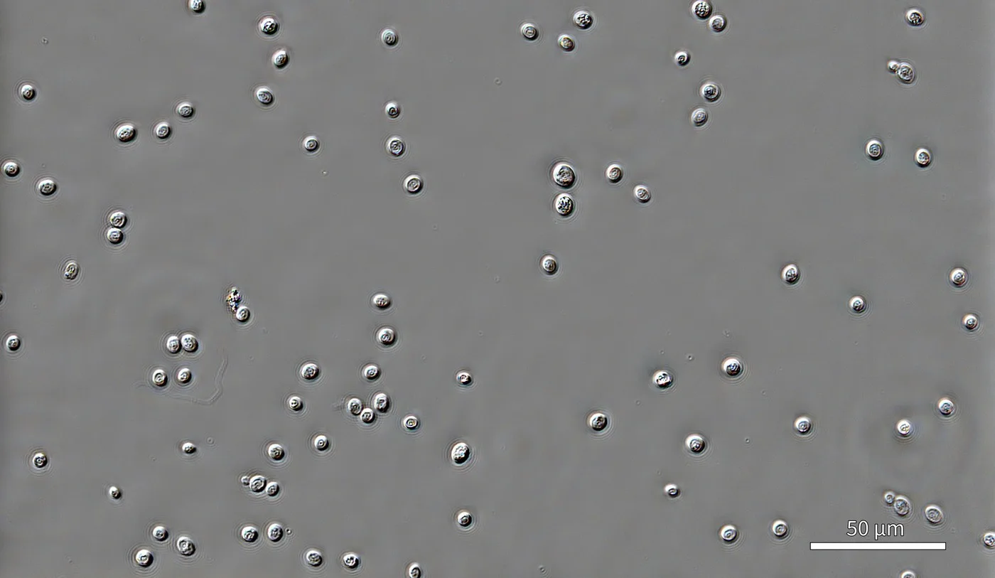
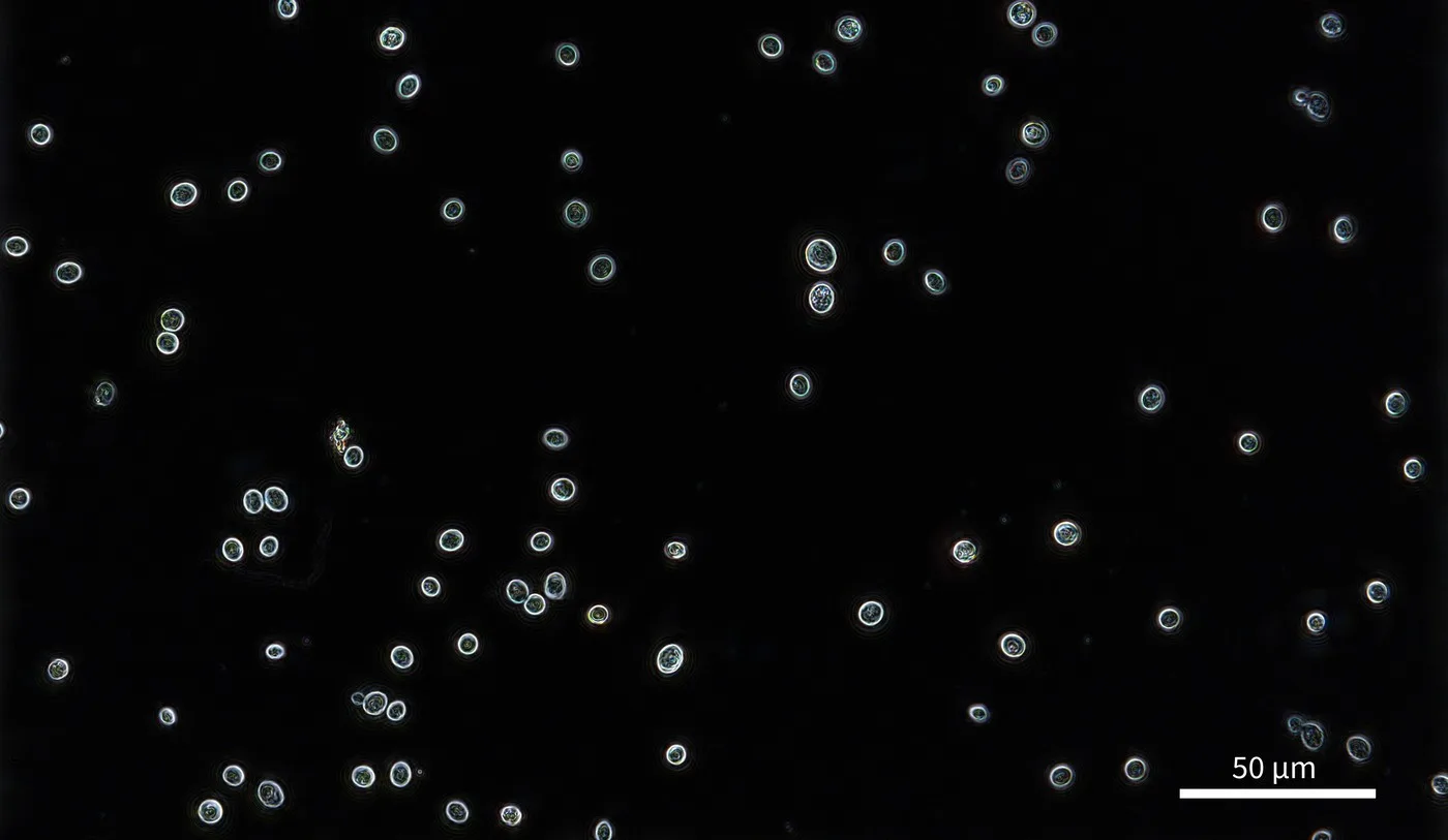
Unstained S. cerevisiae. Scale bar 50 µm.
Dry mass for each cell
With mAIday workflow generator, every cell is segmented and its dry mass read directly from the phase image — no stains, no manual outlining.
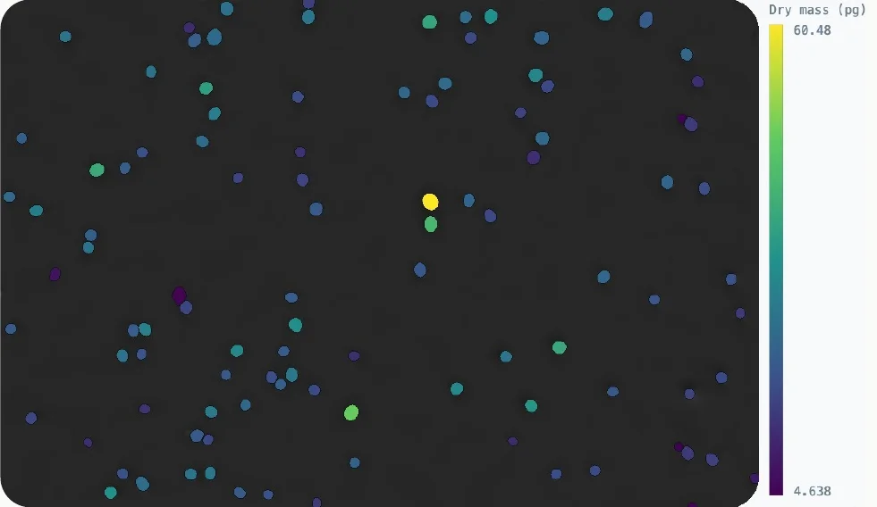
Segmented cells, coloured by dry mass.
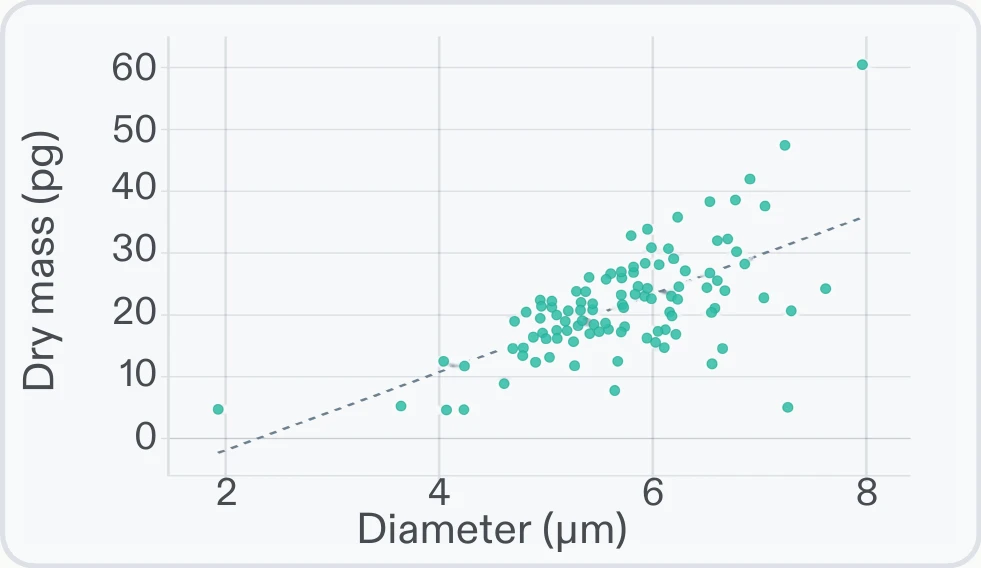
Dry mass vs diameter.
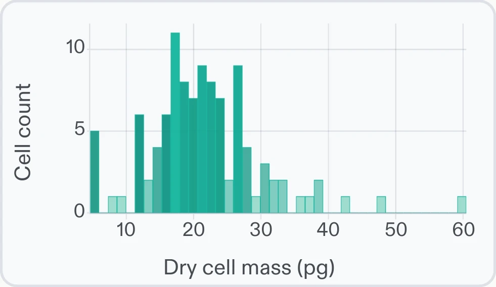
Dry mass distribution (pg).
- 01
Mount and image
No stains, dyes or fixation. Sample prep is loading the labware.
- 02
Dry mass, quantified
Dry cell mass reads directly off the phase image — track growth cell by cell.
- 03
Slide, plate or chip
Slides, multi-well plates and flow chambers — and the Z1 fits inside an incubator.
Organoids · C. elegans · Spheroids · 2D and 3D cell culture · Biocondensates