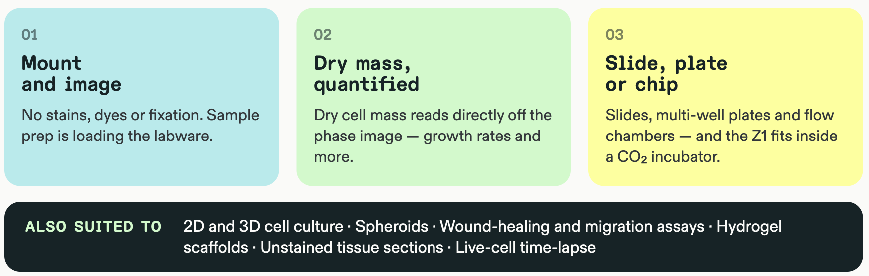
Application
Organoids
Label-free imaging of unstained organoids — no dyes, no fixation, no clearing.
One measurement, four contrast modes
Brightfield for morphology, quantitative phase for dry mass, DIC for fine relief, scattering for boundaries and texture — all from one fast, automated measurement, with the sample left in its culture conditions.
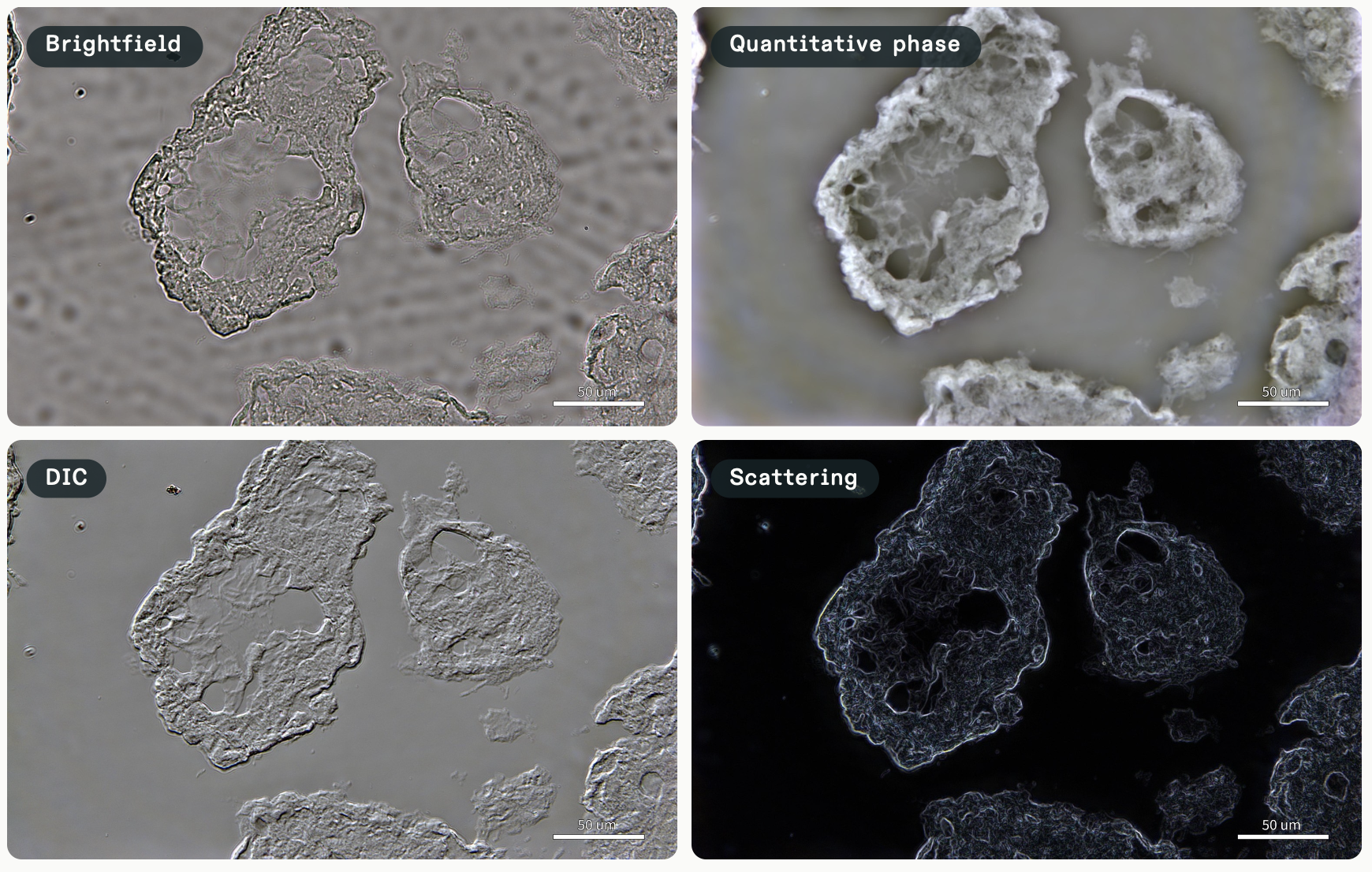
Unstained placental organoids in growth media, slide + coverslip. Sample courtesy of Duke University, USA. Nikon 40× ELWD objective, 5-slice Z-stack ±5 µm, 0.12 µm per pixel. Scale bar 50 µm.