Spores and mycelia
Label-free imaging of live fungal spores and mycelia — no dyes, no fixation, no viability stains.
One measurement, four contrast modes
Brightfield for morphology, quantitative phase for dry mass and internal structure, DIC for fine relief, scattering for edges and texture — all from one fast, automated measurement of live, unstained spores.
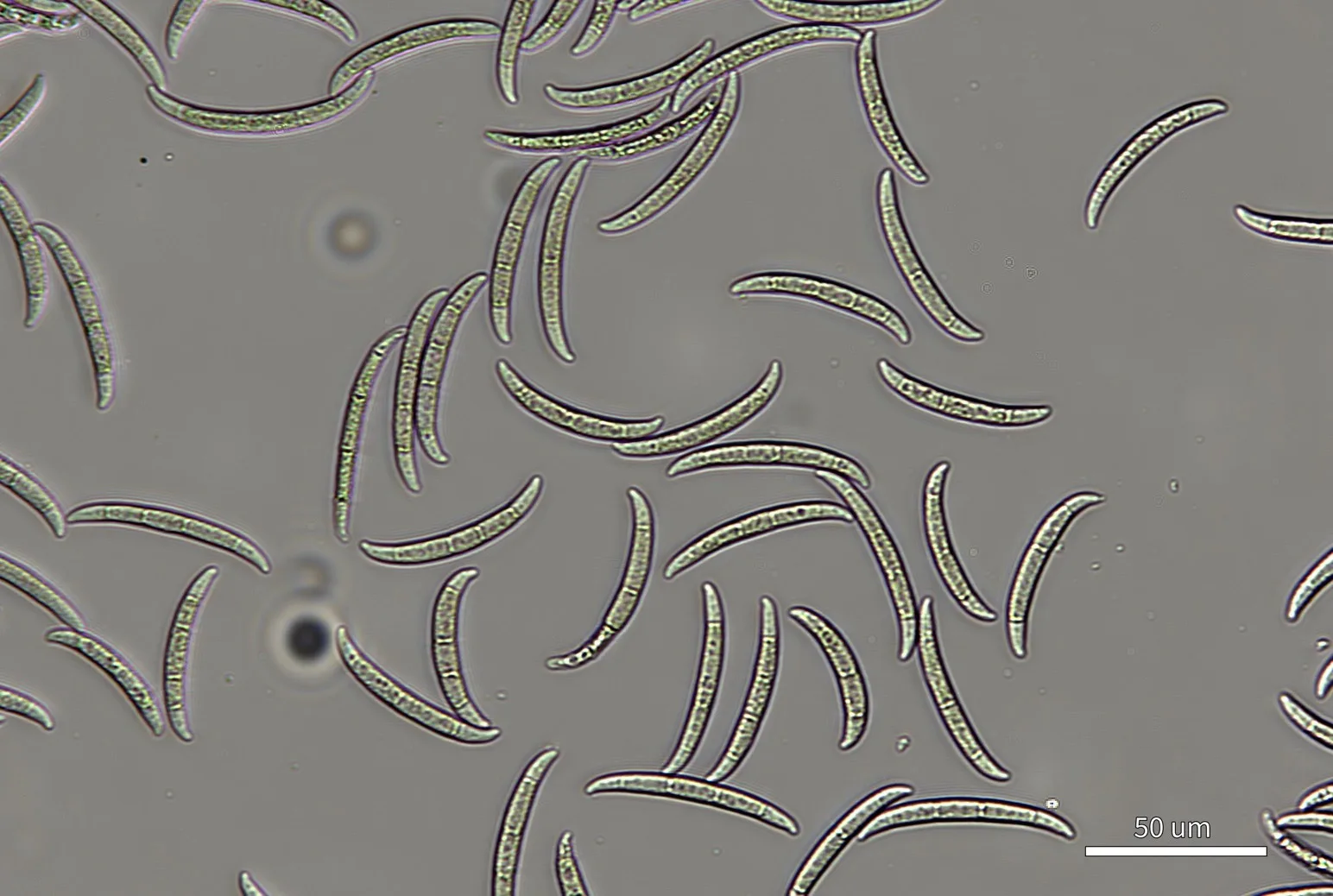
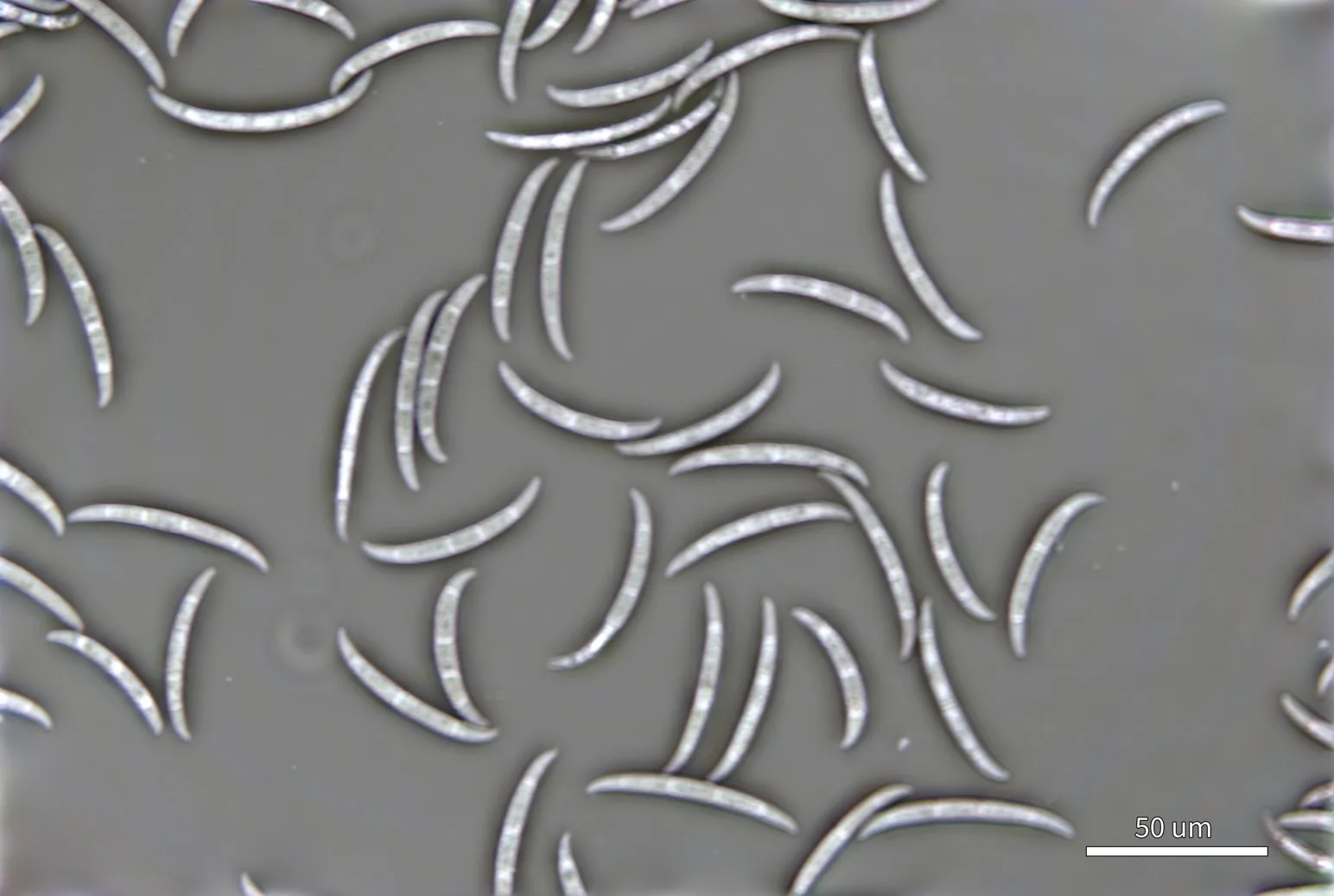
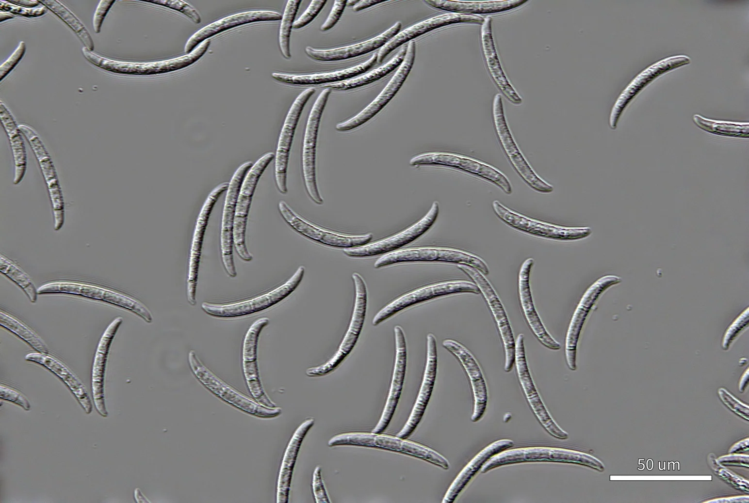
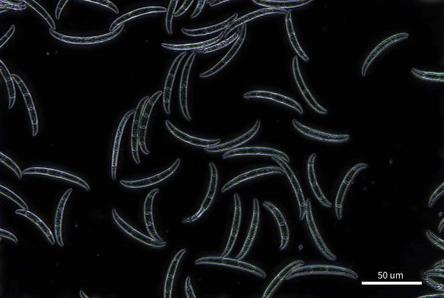
Live, unstained Fusarium sp. spores between coverslips, 130 µm spacer. Sample courtesy of Novonesis, USA. Nikon 40× objective, single frame, 0.12 µm per pixel. Scale bar 50 µm.
- 01
Live or dead, no stain
Dead spores are thinner and internally heterogeneous — a clear phase signature, no viability dye.
- 02
Dry mass, per spore
Automated segmentation and live/dead classification, with dry mass for every spore.
- 03
Spores to mycelia
Z-stacks through 3D mycelia, 384-well plates or coverslips — and multi-well time-lapse scans.
Filamentous fungi and yeast · Spore germination and viability assays · Antifungal screening in multi-well plates · Mycelial growth time-lapse · Fermentation samples
Live and dead, quantified
A population of Fusarium sp. spores was heat-killed and mixed with live spores. The Z1 analysis pipeline then segmented, classified and quantified every spore — end to end on the instrument.
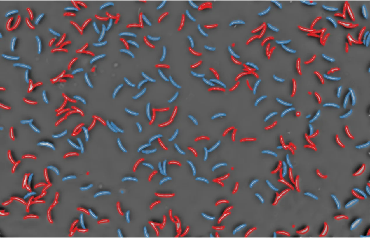
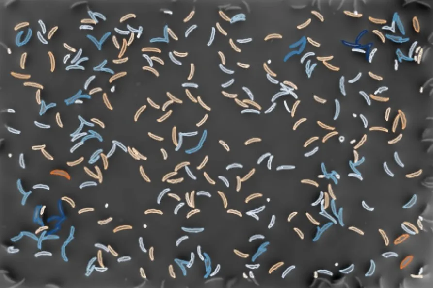
Live and heat-killed Fusarium sp. spores, Nikon 10× objective, 0.48 µm per pixel.
- Every spore, identified. 273 spores segmented and classified in a single field: 136 live and 137 dead.
- Shape and dry mass. Full geometric analysis of each spore — size, shape, projected area and dry mass.
- Automated assays. Histograms and annotated images as output, with automated proliferation and death assays.
Mycelia in 3D
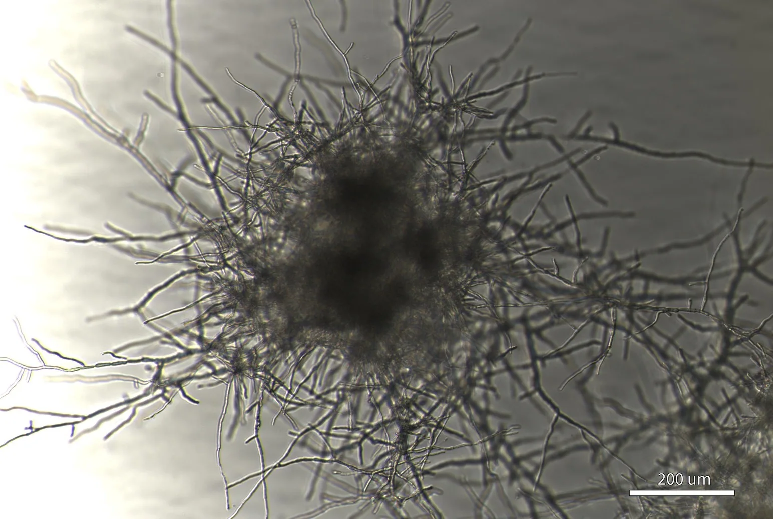
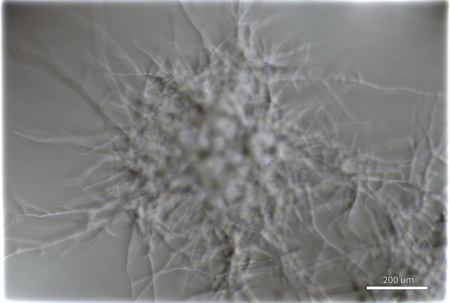
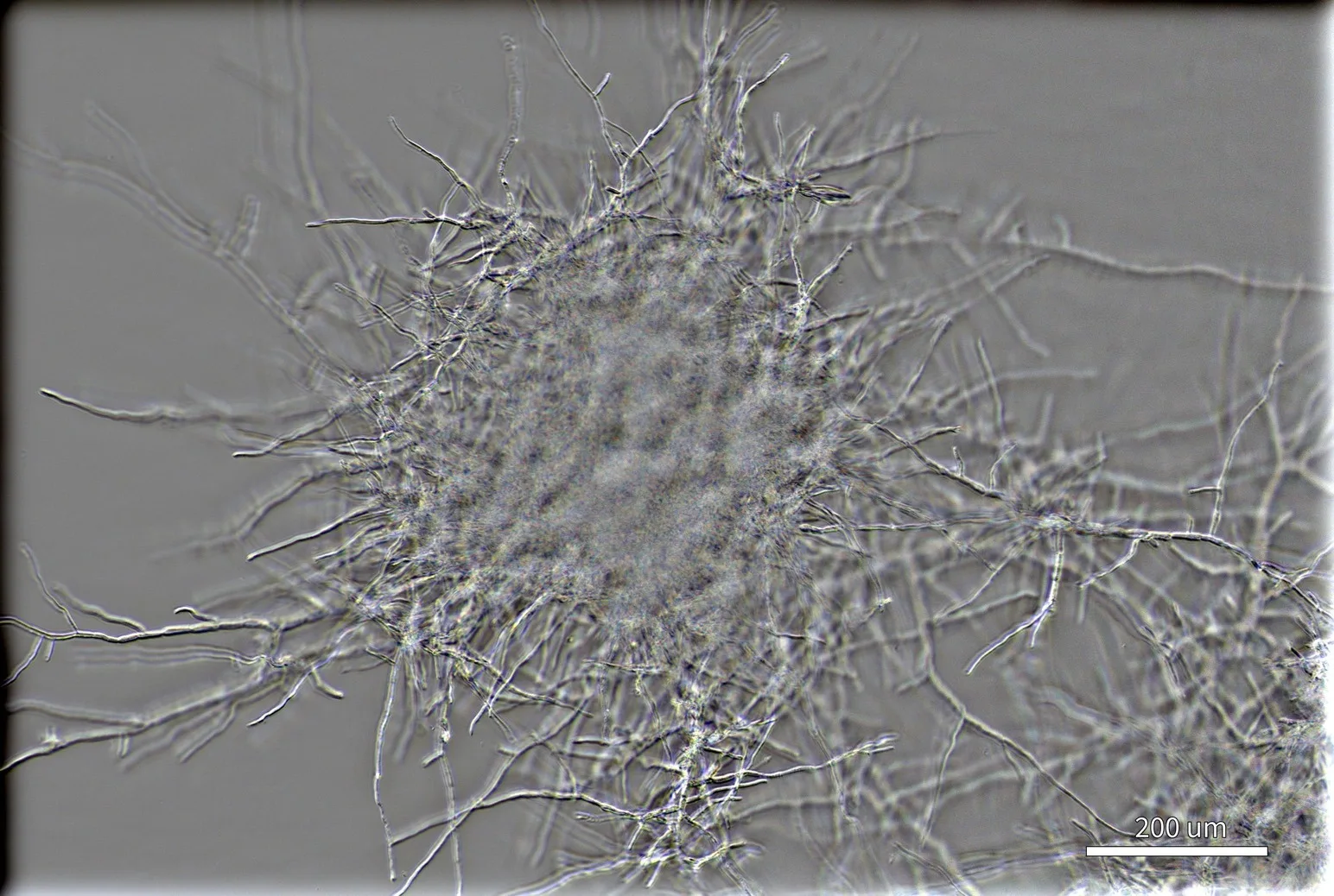
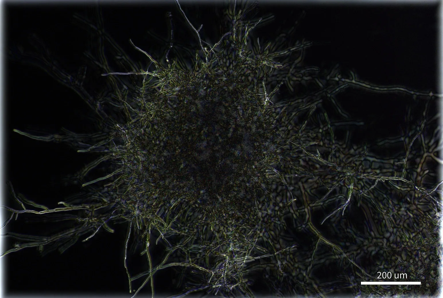
Unstained mycelium, single frame from a 10-step Z-stack. Nikon 10× NA 0.3 objective. Sample courtesy of Novonesis, USA. Scale bar 200 µm.